
Comprehensive hematoma guide covering types, symptoms, causes, and treatments. Learn to recognize dangerous warning signs, when to seek emergency care, and effective recovery strategies.
Introduction

A sudden fall, a hard impact during sports, or even a minor bump can result in more than surface bruising—it can create a hematoma, a potentially serious collection of blood outside blood vessels. While small hematomas often resolve on their own, larger ones or those in critical locations like the brain can become life-threatening emergencies requiring immediate medical intervention. Understanding hematomas—their types, warning signs, risk factors, and treatment options—empowers you to recognize when bruising represents something more serious and when to seek urgent medical care.
What Is a Hematoma? Understanding Blood Pooling
A hematoma occurs when blood vessels rupture and blood collects outside the circulatory system, pooling in surrounding tissues, organs, or body spaces. Unlike bruises (contusions) where blood disperses through tissues, hematomas involve concentrated blood collections forming discrete pockets or masses. The pooled blood may remain liquid initially but often clots, creating firm lumps beneath the skin or within deeper tissues.
How Hematomas Form:
When trauma, surgery, disease, or medication damages blood vessels, blood leaks into surrounding areas. Small vessel injuries typically cause minor bruising, but larger vessel damage produces significant blood accumulation. Your body attempts to contain and reabsorb this blood, but extensive bleeding may overwhelm natural healing processes, requiring medical intervention.
Hematomas range from minor superficial collections resolving spontaneously within weeks to severe internal accumulations requiring emergency surgery. Location, size, and underlying health conditions determine severity and necessary treatment.
Types of Hematomas: Location Matters
Hematomas are classified by their anatomical location, each presenting unique characteristics and risks:
Subcutaneous Hematomas:
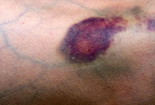
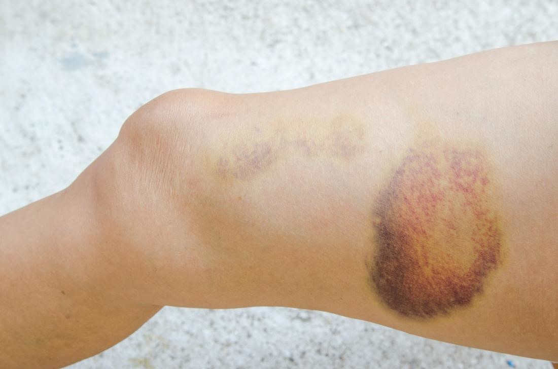
The most common type, occurring just beneath skin surface. These visible hematomas create raised, discolored bumps ranging from small lumps to large masses. Initially appearing red or purple, they transition through green, yellow, and brown as blood breaks down. While painful and unsightly, subcutaneous hematomas rarely cause serious complications.
Intramuscular Hematomas:
Blood collects within muscle tissue, causing deep, aching pain, swelling, and restricted movement. Common in athletes experiencing direct impact or muscle tears. Large intramuscular hematomas may compress nerves or develop compartment syndrome—a dangerous condition requiring emergency treatment.
Subungual Hematomas:
Blood trapped beneath fingernails or toenails from crush injuries or direct trauma. The confined space creates intense, throbbing pressure and pain. The nail typically appears dark purple or black. Drainage often provides significant relief.
Periorbital Hematomas (Black Eyes):
Blood accumulation around eye sockets from facial trauma. While usually not serious, they occasionally indicate underlying skull fractures or brain injury requiring evaluation.
Ear Hematomas (Auricular Hematomas):
Blood collection between ear cartilage and skin, commonly from contact sports or trauma. Untreated ear hematomas can cause permanent deformity (“cauliflower ear”).
Subdural Hematomas:
Life-threatening blood collections between brain and dura (outer brain covering). These develop from head trauma tearing bridging veins. Subdural hematomas may appear immediately (acute) or develop gradually over weeks (chronic), particularly in elderly individuals or those taking blood thinners. Symptoms include headache, confusion, drowsiness, slurred speech, weakness, and seizures.
Epidural Hematomas:
Blood accumulating between skull and dura from arterial bleeding, typically following skull fractures. These create rapidly expanding masses requiring emergency surgery. Classic presentation involves brief unconsciousness, a “lucid interval” of apparent recovery, then rapid deterioration as bleeding continues.
Intracerebral Hematomas:
Bleeding within brain tissue itself, often from severe head trauma or ruptured blood vessels. These cause stroke-like symptoms and require immediate emergency care. Similar to complications seen in cardiovascular disease, intracerebral bleeding can be life-threatening.
Intra-abdominal Hematomas:
Blood collections within abdominal organs or spaces from trauma, surgery, or ruptured blood vessels. May occur in liver, spleen, kidneys, or abdominal cavity. Large intra-abdominal hematomas cause pain, distension, and potentially life-threatening blood loss.
Retroperitoneal Hematomas:
Bleeding in the space behind the abdominal cavity containing kidneys, aorta, and other structures. Often results from trauma, anticoagulation complications, or medical procedures. Can cause massive blood loss without obvious external signs.
Recognizing Hematoma Symptoms: Warning Signs
Symptoms vary dramatically based on hematoma location and size:
Superficial Hematoma Symptoms:
- Visible swelling or raised lump
- Skin discoloration (red, purple, blue, green, yellow, brown progression)
- Pain and tenderness at injury site
- Warmth over affected area
- Limited mobility if near joints
Deep or Internal Hematoma Symptoms:
- Deep, aching pain
- Swelling not visible externally
- Decreased range of motion
- Numbness or tingling from nerve compression
- Muscle weakness
Brain Hematoma Warning Signs (EMERGENCY):
- Severe, worsening headache
- Confusion or altered consciousness
- Drowsiness or difficulty staying awake
- Slurred speech or difficulty speaking
- Vision changes or unequal pupils
- Weakness or numbness on one side of body
- Seizures
- Nausea and vomiting
- Loss of balance or coordination
- Memory problems
Abdominal Hematoma Symptoms:
- Abdominal pain or tenderness
- Swelling or distension
- Bruising on abdomen or flanks
- Dizziness or lightheadedness from blood loss
- Rapid heart rate
- Low blood pressure
Recognizing these symptoms, particularly neurological warning signs, is as critical as identifying symptoms of serious blood disorders—immediate medical attention can save lives.
Causes and Risk Factors: Why Hematomas Develop
Trauma and Injury:
The most common cause involves direct impact, falls, motor vehicle accidents, sports injuries, or assault. Severity depends on force magnitude and vulnerable structures affected.
Medications:
Anticoagulants (Blood Thinners): Warfarin, heparin, apixaban, rivaroxaban, and other anticoagulants prevent clotting, increasing bleeding risk. Even minor trauma can cause significant hematomas.
Antiplatelet Drugs: Aspirin, clopidogrel, and similar medications reduce platelet function, prolonging bleeding after injury.
NSAIDs: Ibuprofen, naproxen, and other non-steroidal anti-inflammatory drugs interfere with platelet function.
Medical Procedures:
Surgery, injections, catheter placements, biopsies, and other procedures may cause iatrogenic hematomas despite careful technique.
Blood Disorders:
Conditions affecting clotting increase hematoma risk:
- Hemophilia (clotting factor deficiencies)
- Von Willebrand disease
- Thrombocytopenia (low platelet counts)
- Liver disease affecting clotting protein production
Blood Vessel Abnormalities:
Aneurysms, arteriovenous malformations, or fragile vessels from aging rupture spontaneously or with minimal trauma.
Age:
Elderly individuals experience hematomas more frequently due to fragile blood vessels, thinner skin, and frequent anticoagulant use. Falls become increasingly dangerous with age.
Excessive Alcohol Use:
Chronic alcohol consumption damages liver function, reducing clotting factor production and increasing bleeding risk.
Diagnosis: How Doctors Evaluate Hematomas
Physical Examination:
Doctors assess visible hematomas, noting size, location, tenderness, and associated symptoms. Neurological examination detects brain hematoma signs.
Imaging Studies:
Ultrasound: Evaluates superficial and some deep hematomas, distinguishing them from other masses.
CT Scan: The primary tool for detecting brain hematomas, showing blood location, size, and effects on surrounding structures. Also evaluates abdominal and pelvic hematomas.
MRI: Provides detailed soft tissue images, particularly useful for older hematomas or complex cases.
X-rays: Detect associated fractures that may have caused hematomas.
Laboratory Tests:
Complete Blood Count: Assesses blood loss severity and platelet counts.
Coagulation Studies: Measure clotting function (PT, PTT, INR), identifying bleeding disorders or medication effects.
Blood Chemistry: Evaluates organ function and electrolyte balance.
According to the Cleveland Clinic, prompt diagnosis of internal hematomas, particularly in the brain, is crucial for preventing permanent disability or death.
Treatment Options: Managing Hematomas Effectively
Treatment depends on hematoma location, size, and symptoms:
Conservative Management (Small, Uncomplicated Hematomas):
Rest: Avoid activities that might worsen bleeding or reinjure the area.
Ice Application: Apply ice packs for 15-20 minutes several times daily during the first 48 hours, reducing swelling and pain.
Compression: Elastic bandages minimize swelling and provide support for extremity hematomas.
Elevation: Raising injured areas above heart level reduces swelling and promotes drainage.
Pain Management: Acetaminophen for pain (avoid aspirin and NSAIDs that promote bleeding).
Medical Interventions:
Drainage (Aspiration): Using needles or small incisions to remove accumulated blood, providing relief from pressure and pain. Common for subungual, ear, and some superficial hematomas.
Surgical Evacuation: Large or expanding hematomas, particularly those causing organ compression or neurological symptoms, require surgical removal. Brain hematomas often need emergency craniotomy to evacuate blood and relieve pressure.
Embolization: Minimally invasive procedure where interventional radiologists block bleeding vessels using coils, particles, or adhesives, treating ongoing hemorrhage without surgery.
Blood Transfusions: For significant blood loss causing anemia or hemodynamic instability.
Reversal Agents: Medications reversing anticoagulant effects, stopping ongoing bleeding in patients taking blood thinners.
Treatment by Location:
Brain Hematomas: Require emergency neurosurgical evaluation. Treatment ranges from observation with close monitoring to urgent surgical evacuation depending on size, location, and symptoms. Similar to managing chronic neurological conditions, specialized care is essential.
Subungual Hematomas: Nail trephination (creating small hole to drain blood) relieves pressure and pain.
Ear Hematomas: Drainage plus compression dressing prevents cartilage damage and deformity.
Muscle Hematomas: Rest, ice, compression, elevation (RICE protocol) with gradual return to activity guided by physical therapy.
Recovery and Healing: What to Expect
Healing Timeline:
- Small superficial hematomas: 2-4 weeks
- Moderate hematomas: 4-8 weeks
- Large deep hematomas: Several months
- Brain hematomas: Variable; depends on severity and complications
Color Changes:
As blood breaks down, hematomas transition through characteristic color progression: red/purple → blue → green → yellow → brown, eventually fading completely.
Potential Complications:
Infection: Particularly with open drainage or large hematomas. Signs include increasing pain, warmth, redness, fever, and pus drainage.
Compartment Syndrome: Pressure buildup within muscle compartments restricting blood flow, causing permanent tissue damage. Requires emergency fasciotomy (surgical pressure release).
Calcification (Myositis Ossificans): Calcium deposits forming within healing hematomas, creating hard masses. Common in thigh and arm muscles.
Chronic Subdural Hematomas: Ongoing brain compression requiring delayed surgical intervention.
Scarring: Large hematomas may leave permanent discoloration, indentation, or lumps.
Prevention Strategies: Reducing Hematoma Risk
Safety Measures:
- Wear protective equipment during sports (helmets, pads, guards)
- Use handrails on stairs
- Remove tripping hazards at home
- Install adequate lighting
- Wear appropriate footwear
- Use assistive devices if needed for mobility
Medication Management:
If taking anticoagulants or antiplatelets:
- Follow dosing instructions precisely
- Attend regular monitoring appointments
- Inform all healthcare providers about medications
- Avoid unnecessary NSAIDs or supplements affecting bleeding
- Report unusual bruising or bleeding promptly
Fall Prevention:
Particularly important for elderly individuals taking blood thinners:
- Regular vision and hearing checks
- Balance and strength exercises
- Home safety modifications
- Review medications affecting balance
- Adequate preventive care addressing fall risks
Managing Underlying Conditions:
Control bleeding disorders, maintain liver health, and address conditions increasing hematoma risk through comprehensive medical management.
Frequently Asked Questions About Hematomas
How can I tell if a hematoma is serious?
Seek immediate medical attention if hematomas occur after head trauma with symptoms like severe headache, confusion, vomiting, or loss of consciousness. Large hematomas causing significant swelling, severe pain, numbness, or movement restriction require evaluation. Hematomas in people taking blood thinners, those with bleeding disorders, or bleeding not stopping with pressure need medical assessment. Trust your instincts—when concerned about severity, seek professional evaluation.
Can hematomas cause permanent damage?
Most superficial hematomas heal completely without lasting effects. However, complications can cause permanent damage: brain hematomas may cause neurological deficits, compartment syndrome can destroy muscles and nerves, large muscle hematomas may calcify creating permanent lumps, and untreated ear hematomas cause permanent deformity. Prompt appropriate treatment minimizes permanent damage risk.
Should I apply heat or ice to a hematoma?
Apply ice during the first 48-72 hours after injury to reduce swelling, pain, and further bleeding. Use ice packs for 15-20 minutes several times daily. After 2-3 days, switch to heat application promoting blood flow and reabsorption of pooled blood. Warm compresses or heating pads for 15-20 minutes several times daily accelerate healing during later recovery stages.
How long does it take for a hematoma to go away?
Small superficial hematomas typically resolve within 2-4 weeks. Moderate-sized hematomas may take 4-8 weeks, while large deep hematomas can require several months for complete resolution. Brain hematomas vary significantly—some small ones reabsorb over weeks to months, while others require surgical evacuation. Factors affecting healing include hematoma size, location, your age, overall health, and whether complications develop.
Can I exercise with a hematoma?
Rest is crucial initially to prevent worsening bleeding and allow healing to begin. Avoid activities involving the affected area for at least several days to weeks depending on severity. Gradually resume gentle movement and exercise as pain and swelling decrease, following healthcare provider or physical therapist guidance. Premature return to vigorous activity risks reinjury, bleeding recurrence, or complications. Brain hematoma patients require specific medical clearance before resuming physical activities.
Take Hematomas Seriously: Know When to Act
While many hematomas represent minor injuries healing without intervention, some become medical emergencies requiring immediate attention. Understanding the difference between benign bruising and dangerous blood collections empowers you to respond appropriately, potentially preventing disability or saving lives.
Never dismiss concerning symptoms, particularly after head trauma or in individuals taking anticoagulants. When in doubt about hematoma severity, seek medical evaluation—early intervention prevents complications and ensures optimal outcomes. Maintain awareness of your body’s healing progress, report unexpected changes to healthcare providers, and follow treatment recommendations carefully.
Your vigilance in recognizing warning signs, combined with prompt appropriate medical care when needed, provides the best protection against serious hematoma complications. Stay informed, stay safe, and prioritize your health by taking all injuries seriously until proven minor.
If you’ve experienced significant trauma, notice unusual bleeding or bruising, or develop concerning symptoms, don’t wait—seek medical evaluation immediately. Your health and safety are too important to risk.