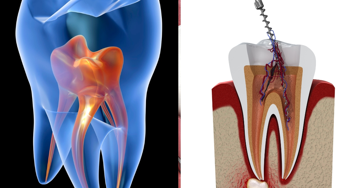

Discover the vital role of tooth pulp – the living core of your tooth. Learn its functions, what causes painful infections, and how root canal treatment saves your smile.
Introduction: The Beating Heart Within Your Smile
Deep within the hardest substance in your body lies its most vulnerable and vital core: the dental pulp. Often thought of only when it screams in pain from a deep cavity, the pulp is far more than a simple nerve. It is the dynamic, living engine of your tooth—a complex tissue responsible for its creation, nourishment, sensation, and defense. Understanding the pulp is key to appreciating why a tooth is a living organ, not a static stone, and why its health is paramount to your smile’s survival. This guide will take you on a journey into the very heart of your tooth, exploring the pulp’s remarkable anatomy, its critical functions, and the dramatic consequences when it becomes diseased, empowering you with knowledge to protect this hidden cornerstone of oral health.
What is the Dental Pulp? Anatomy of a Living Core
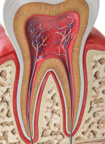
The dental pulp is the soft, living connective tissue located in the central hollow space of a tooth. This space is called the pulp chamber (in the crown) and the root canal(s) (extending down the root). Far from being a blank cavity, it is a densely packed, highly organized structure often described as the “tooth’s vital organ.”
Think of a tooth like a house:
- Enamel: The hard, shiny, white roof and walls.
- Dentin: The sturdy, bony framework underneath the enamel, making up the bulk of the tooth.
- Pulp: The living family and utilities inside—the electrical wiring (nerves), plumbing (blood vessels), and repair crew (cells) that keep the house functional and responsive to the environment.
The Multi-Layered Structure of the Pulp
The pulp is not homogeneous. From its outer walls inward, it consists of distinct zones:
- Odontoblast Layer: The pulp’s first line of defense. These specialized cells line the outer wall, directly against the dentin. They are famous for producing dentin throughout the tooth’s life. Each odontoblast has a long cytoplasmic process that extends into a microscopic tubule in the dentin, creating a direct biological connection between the pulp and the hard structure of the tooth.
- Cell-Free Zone (of Weil): A relatively clear zone beneath the odontoblasts, rich in nerve and capillary networks.
- Cell-Rich Zone: This area contains a high density of fibroblasts (the pulp’s main structural cells) and undifferentiated mesenchymal stem cells, which are crucial for repair.
- Pulp Core: The central region containing the tooth’s major blood vessels and nerves, which enter and exit through a tiny opening at the tip of the root (the apex).
The Pulp’s Cellular Workforce
The pulp’s functions are carried out by a dedicated team of cells:
- Odontoblasts: The dentin factories. They create primary dentin during tooth formation and secondary dentin slowly throughout life, which narrows the pulp chamber. They can also rapidly produce reparative (tertiary) dentin in response to injury like decay or wear, acting as a natural barrier to shield the pulp.
- Fibroblasts: The maintenance crew. They produce and maintain the pulp’s extracellular matrix (the collagenous scaffold that gives the tissue its structure).
- Immune Cells (Macrophages, Lymphocytes, etc.): The defense force. They patrol for and attack invading bacteria.
- Undifferentiated Mesenchymal Cells: The regenerative reserve. These stem cells can transform into odontoblasts or other cells needed for repair, representing the pulp’s remarkable healing potential.
- Nerves and Blood Vessels: The communication and supply lines. A rich network provides sensation and delivers vital oxygen and nutrients.
Table 1: The Cellular Inhabitants of the Dental Pulp
| Cell Type | Primary Role | Why It Matters |
|---|---|---|
| Odontoblasts | Produce and maintain dentin. | Forms the tooth’s bulk; creates reparative dentin to wall off injury. |
| Fibroblasts | Synthesize the pulp’s structural matrix (collagen). | Maintains the pulp’s healthy connective tissue framework. |
| Immune Cells | Defend against bacterial invasion. | Initiates inflammation to fight infection; part of the pulp’s immune response. |
| Stem Cells | Serve as a regenerative reservoir. | Can differentiate into odontoblasts for repair; focus of cutting-edge research. |
| Nerves & Vessels | Provide sensation and nutrient/waste transport. | Allows tooth to sense stimuli; keeps pulp tissue alive and hydrated. |
The Critical Functions of the Pulp: More Than Just Pain
The pulp is not a passive occupant. It performs several indispensable functions that are categorized as formative, nutritive, sensory, and defensive.
1. Formative (Dentinogenesis)
This is the pulp’s fundamental job. The odontoblasts create all the dentin in the tooth. Without a healthy pulp during development, a tooth simply would not form.
2. Nutritive
The extensive network of blood vessels (both arteries and veins) within the pulp supplies water, oxygen, glucose, amino acids, and minerals to the surrounding dentin and the odontoblasts. This keeps the dentin hydrated and the overall tooth structure resilient. Think of dentin as a sponge that needs to stay moist; a non-vital (dead) tooth becomes brittle and more prone to fracture over time.
3. Sensory (Innervation)
The pulp is densely innervated by both myelinated A-delta fibers and unmyelinated C-fibers.
- A-delta fibers: Respond quickly to stimuli like cold, air, or drilling, producing a sharp, sudden pain. This is your early warning system.
- C-fibers: Respond to deeper inflammation, pressure, and heat, producing a dull, throbbing, and often unbearable ache associated with advanced pulpitis (pulp inflammation).
This sensory function is protective. Pain is a signal that something is wrong, prompting you to seek care.
4. Defensive & Reparative
This is perhaps the pulp’s most sophisticated capability. When threatened—most commonly by bacterial toxins from tooth decay—the pulp mounts a complex response.
- Inflammatory Response: Blood vessels dilate, immune cells rush to the site, and the pulp tissue swells. This is pulpitis, which causes the classic toothache.
- Reparative Dentinogenesis: The odontoblasts are stimulated to rapidly lay down a new, irregular layer of reparative (tertiary) dentin on the wall of the pulp chamber facing the injury. This acts like a wall of scar tissue, attempting to wall off the advancing decay and protect the pulp’s vitality.
The success of this defense determines whether the pulp survives (reversible pulpitis) or succumbs to necrosis (irreversible pulpitis).
When the Pulp is in Peril: Disease and Consequences
The pulp’s greatest enemy is bacteria. Its location inside a rigid, unyielding chamber of dentin and enamel is both its sanctuary and its prison.
The Pathway to Infection
- Deep Decay: Untreated dental caries (cavities) progress through the enamel and into the dentin. The bacteria and their acidic byproducts travel down the dentinal tubules—the very pathways created by the odontoblast processes.
- Pulpitis: The bacterial invasion triggers inflammation (pulpitis). In the early stage (reversible pulpitis), the pulp is irritated but can heal if the decay is removed and a filling is placed. The hallmark symptom is sharp pain to cold or sweets that subsides quickly.
- Irreversible Pulpitis: If the insult is too severe or prolonged, the inflammation overwhelms the pulp’s defenses. The swelling increases, but because the pulp is trapped within rigid tooth walls, the pressure builds, crushing its own blood vessels and cutting off its blood supply. This causes severe, spontaneous, throbbing pain, often worsened by heat and lying down. The pulp cannot recover at this stage; it is dying.
- Pulp Necrosis: The pulp tissue dies. The pain may suddenly stop as the nerves die, giving a false sense of relief. However, the bacteria now have a necrotic tissue to feast on and multiply within the root canal system.
- Apical Periodontitis: The infection and bacterial toxins exit through the apex (tip) of the root, inflaming the surrounding bone and ligament. This can lead to a dental abscess—a pus-filled pocket at the root tip causing pain on biting, swelling, and potentially systemic illness.
Other Causes of Pulp Damage
- Trauma: A physical blow can sever the pulp’s blood supply at the apex.
- Cracks/Fractures: A deep crack can provide a direct pathway for bacteria.
- Severe Wear or Erosion: From grinding (bruxism) or acid reflux, which can wear through enamel and dentin.
- Aggressive Dental Procedures: Repeated deep fillings or preparations for crowns can sometimes irritate the pulp.
Saving the Tooth: Endodontic Treatment (Root Canal Therapy)
When the pulp is irreversibly inflamed or necrotic, the treatment of choice is endodontic therapy, or a root canal, performed by a general dentist or an endodontist (a root canal specialist). The goal is not to “kill the nerve,” but to remove the diseased tissue, disinfect the canal space, and seal it to preserve the tooth.
The Modern Root Canal Process:
- Diagnosis: Using symptoms, clinical tests (cold, electric pulp tester, percussion), and X-rays to assess pulp status.
- Anesthesia and Isolation: The tooth is numbed, and a rubber dam is placed to keep it clean and saliva-free.
- Access Opening: A small opening is made in the crown to reach the pulp chamber.
- Cleaning and Shaping: Using tiny instruments, the infected pulp tissue is removed from the chamber and root canals. The canals are then cleaned, disinfected, and shaped to receive a filling.
- Filling and Sealing: The empty, clean canals are filled with a biocompatible material called gutta-percha and a sealer to prevent re-infection.
- Final Restoration: The access opening is sealed, and a crown is usually recommended to restore full strength and function.
Contrary to popular myth, a properly root-canaled tooth is not “dead.” It is a non-vital tooth (lacking a living pulp) but remains a functional, living part of your jawbone, held in place by the healthy periodontal ligament. It can last a lifetime with proper care.
The Future: Regenerative Endodontics and Pulp Preservation
The frontier of endodontics aims to move beyond simply removing diseased pulp to regenerating healthy tissue. Regenerative endodontics leverages the pulp’s own stem cells. The concept involves disinfecting the root canal system and then introducing scaffolds and growth factors to encourage stem cells to differentiate into new, functional pulp-like tissue, potentially allowing for continued tooth development and natural defense in young patients. This represents a paradigm shift from disease management to true biological restoration.
FAQs: Your Questions About Tooth Pulp Answered
1. Why does pulp pain hurt so much?
The pulp is an exquisitely sensitive tissue encased in a rigid compartment. When it becomes inflamed and swells, the pressure has nowhere to go. This pressure buildup compresses the nerves, causing intense pain. The types of nerve fibers (C-fibers) activated in deep inflammation are also associated with the most severe, throbbing pain sensations in the body.
2. If my tooth stops hurting, does that mean the infection is gone?
No. This is a dangerous misconception. The sudden cessation of pain often means the pulp has fully necrosed (died). The nerves that carry pain signals are no longer functional. However, the bacteria are still present and actively multiplying, and the infection is now spreading into the bone around the root tip, which can eventually cause a painful abscess and bone loss. Always see a dentist for persistent tooth pain, even if it goes away.
3. Can a damaged pulp heal on its own?
A mildly irritated pulp (reversible pulpitis) can heal if the source of irritation (like decay) is completely removed and the tooth is properly restored. However, once the pulp reaches a state of irreversible inflammation or infection, it cannot heal itself. The damage is progressive and will lead to necrosis without professional intervention.
4. Is it better to have a root canal or just pull the tooth?
Preserving your natural tooth via root canal therapy is almost always the recommended course. No artificial replacement—bridge, implant, or denture—matches the function and efficiency of a natural tooth. Root canal treatment has a very high success rate (over 95%) and allows you to keep your natural tooth structure, maintaining jawbone health and proper chewing function. Extraction should be a last resort. For more on replacement options, see our guide on types of dental treatments.
5. How can I keep my tooth pulp healthy?
The best strategy is prevention:
- Maintain excellent oral hygiene to prevent deep decay.
- Use fluoride toothpaste to strengthen enamel and dentin.
- Wear a mouthguard during sports to prevent traumatic injury.
- Address teeth grinding (bruxism) with a night guard.
- See your dentist regularly for check-ups to catch and treat cavities while they are still small and far from the pulp.
Conclusion: Honoring the Life Within Your Tooth
The dental pulp is a masterpiece of biological engineering—a tiny, dynamic world responsible for creating, sustaining, and defending your teeth. It is the reason your teeth can sense an ice cube, adapt to wear, and warn you of danger. Understanding this hidden “life source” transforms your view of a tooth from a static object to a living, responsive part of your body. By prioritizing preventive care and seeking prompt treatment for dental problems, you honor this complex tissue and invest in the long-term health and preservation of your natural smile. Remember, every toothache is a message from the pulp; listening to it early is the key to saving it.