
Comprehensive tooth structure guide covering anatomy layers, functions, protection strategies, and essential dental health insights for maintaining strong teeth.
Introduction
Your teeth are remarkable biological structures—miniature engineering marvels designed to last a lifetime with proper care. Understanding tooth structure and anatomy empowers you to protect these irreplaceable assets, recognize when problems develop, and make informed decisions about dental care. Each tooth contains multiple specialized layers working together to enable biting, chewing, speaking, and supporting facial structure. This comprehensive guide explores the intricate architecture of teeth, explains how each component functions, and provides powerful strategies for preserving dental health throughout your life.
Overview of Tooth Anatomy

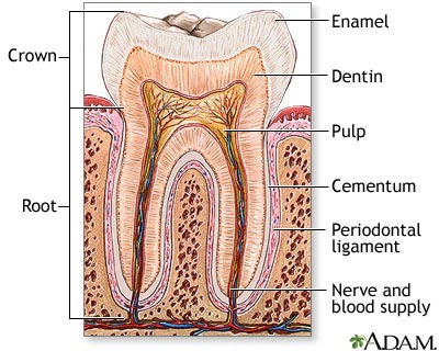
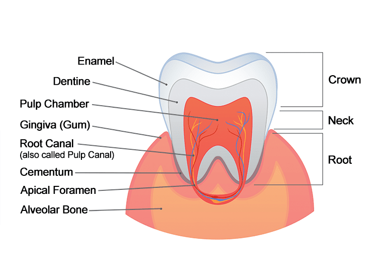
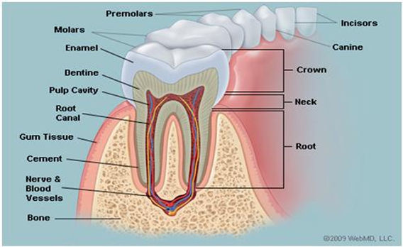
A tooth consists of two main regions: the crown (visible portion above the gum line) and the root (hidden portion anchored in the jawbone). Each tooth comprises four primary layers—enamel, dentin, cementum, and pulp—plus supporting structures that hold teeth firmly in place.
Understanding these components helps you appreciate why certain dental problems occur and how treatments work. While teeth appear simple from the outside, their internal complexity rivals many other body structures in sophistication and functionality.
The Four Primary Layers of a Tooth
Enamel: The Protective Outer Shell
Enamel forms the outermost layer covering the tooth crown—the portion visible in your mouth. This remarkable substance is the hardest tissue in the human body, even harder than bone. Enamel’s extreme hardness enables it to withstand tremendous biting and chewing forces that can exceed 200 pounds per square inch.
Composition and Structure: Enamel consists of approximately 96 percent minerals (primarily hydroxyapatite crystals containing calcium and phosphate), 3 percent water, and 1 percent organic material. This high mineral content creates the dense, crystalline structure responsible for enamel’s exceptional hardness and translucent appearance.
Enamel crystals are organized into rod-like structures (enamel prisms) running from the tooth’s inner layers toward the surface. This organized architecture maximizes strength while allowing slight flexibility to absorb shock during chewing.
Functions: Enamel serves multiple critical purposes including protecting underlying tooth structures from damage, resisting wear from biting and chewing, preventing bacterial penetration that would cause decay, insulating teeth from temperature extremes (hot and cold foods), and providing the smooth, white appearance of healthy teeth.
Vulnerabilities: Despite its hardness, enamel is vulnerable to acid erosion from dietary acids, bacterial acid produced during tooth decay, wear from grinding or clenching teeth, and damage from trauma or using teeth as tools. Once damaged, enamel cannot regenerate—the body cannot produce new enamel after tooth formation. This makes protection crucial.
Dentin: The Supportive Core
Beneath the enamel lies dentin, which forms the bulk of the tooth structure. Dentin extends throughout both the crown and root, comprising the majority of tooth mass. While harder than bone, dentin is softer than enamel and has a yellowish color that influences overall tooth appearance.
Composition and Structure: Dentin consists of approximately 70 percent minerals, 20 percent organic material (primarily collagen), and 10 percent water. This composition makes dentin more resilient and flexible than brittle enamel.
Dentin contains millions of microscopic tubules (tiny channels) running from the pulp cavity to the outer dentin boundary. These tubules contain fluid and nerve extensions from the pulp, making dentin a living, sensitive tissue unlike inert enamel.
Functions: Dentin provides structural support for overlying enamel, transmits sensory information from stimuli to nerve tissue in the pulp, protects the pulp from external damage and temperature changes, and continues forming throughout life in response to stimuli (secondary and tertiary dentin).
Sensitivity: The tubules in dentin make this layer sensitive to temperature, pressure, and chemical stimuli. When enamel wears away or gums recede exposing dentin, sensitivity occurs as stimuli reach nerve endings through the tubules. This sensitivity is why exposed dentin causes discomfort with hot, cold, sweet, or acidic foods.
Cementum: The Root Covering
Cementum is a specialized calcified tissue covering the tooth root—the portion normally hidden below the gum line. Cementum is similar to bone in composition but lacks the blood vessels and nerve supply that bone contains.
Composition and Structure: Cementum consists of approximately 45-50 percent minerals, making it softer than both enamel and dentin. It contains collagen fibers that anchor into the periodontal ligament—the connective tissue holding teeth in their sockets.
Functions: Cementum’s primary purpose is anchoring the tooth to the jawbone via the periodontal ligament. Collagen fibers from the periodontal ligament embed in cementum on one end and jawbone on the other, creating a strong yet flexible attachment that allows slight tooth movement to absorb chewing forces.
Cementum also continues forming throughout life, adapting to changing forces and compensating for natural tooth wear by lengthening roots slightly.
Exposure Concerns: Gum recession can expose cementum to the oral environment. Since cementum is much softer than enamel, exposed cementum is highly vulnerable to decay, wear, and sensitivity. This makes gum health critical for protecting tooth roots.
Pulp: The Living Core
At the tooth’s center lies the pulp—soft tissue containing blood vessels, nerves, and connective tissue. The pulp occupies the pulp chamber in the crown and extends through root canals down to the root tips (apex).
Composition and Functions: Dental pulp is highly vascularized (rich in blood vessels) and innervated (containing many nerve fibers). The pulp performs several vital functions including supplying nutrients to dentin through blood vessels, providing sensory function allowing you to feel pressure, temperature, and pain, and producing dentin throughout life (dentin formation continues in response to wear, decay, or injury).
During tooth development, pulp is essential for forming the tooth structure. In mature teeth, the pulp becomes less critical—teeth can survive without pulp after root canal treatment, though they become somewhat more brittle and vulnerable to fracture.
Pulp Vulnerability: The pulp is vulnerable to bacterial infection from deep decay, inflammation from trauma or extensive dental work, and degeneration with aging. According to the American Dental Association, understanding tooth anatomy helps people recognize when dental problems require professional attention before they progress to painful pulp infections.
Supporting Structures
Beyond the tooth itself, several surrounding structures provide essential support and protection.
Periodontal Ligament: This network of connective tissue fibers surrounds the tooth root, connecting cementum to the alveolar bone (tooth socket). The periodontal ligament acts as a shock absorber, cushioning teeth during chewing and allowing slight movement that prevents damage from excessive forces. It also contains sensory nerves that help you gauge biting force and position your teeth precisely.
Alveolar Bone: The specialized jawbone that contains tooth sockets (alveoli) holds teeth in position. This bone is living tissue that constantly remodels in response to chewing forces. Loss of teeth leads to bone loss since the stimulation from chewing is removed. Maintaining healthy teeth helps preserve jawbone structure.
Gingiva (Gums): The soft tissue covering the alveolar bone and surrounding tooth necks where crowns meet roots. Healthy gums are firm, pink, and don’t bleed during normal brushing or flossing. Gums protect underlying bone and tooth roots from bacteria and trauma. Gum disease can lead to recession, exposing sensitive root surfaces and eventually causing tooth loss.
Types of Teeth and Their Specialized Functions
Different tooth types in your mouth have specialized shapes suited to particular functions. Understanding this specialization helps explain why different teeth experience different problems.
Incisors: The eight front teeth (four upper, four lower) with sharp, chisel-shaped edges designed for cutting food. Incisors are most visible when smiling and speaking.
Canines: The four pointed teeth flanking the incisors (two upper, two lower) designed for tearing food. Canines have the longest roots of any teeth and are among the last to be lost.
Premolars (Bicuspids): The eight teeth behind canines (four upper, four lower) with broader, flatter surfaces and small points (cusps) for crushing and grinding food.
Molars: The twelve back teeth (six upper, six lower—or eight if wisdom teeth are present) with large, flat surfaces and multiple cusps designed for grinding food. Molars do the heavy work of chewing and are most prone to decay due to their deep grooves that can trap food and bacteria.
Tooth Development and Changes Over Time
Understanding how teeth develop and change throughout life helps you protect them appropriately at different ages.
Primary Teeth (Baby Teeth): Children develop 20 primary teeth beginning around 6 months and completing by age 3. These teeth have thinner enamel than permanent teeth, making them more vulnerable to decay. Primary teeth are crucial for proper jaw development, speech development, and holding space for permanent teeth.
Permanent Teeth: Beginning around age 6, permanent teeth gradually replace primary teeth, with the process typically complete by age 13 (except wisdom teeth, which emerge in late teens or early twenties). Adults have 32 permanent teeth if all teeth including wisdom teeth are present, or 28 without wisdom teeth.
Age-Related Changes: Teeth undergo several changes with aging including enamel thinning from years of wear, dentin darkening causing teeth to appear more yellow, pulp shrinking (reducing sensitivity but also reducing regenerative capacity), and increased brittleness. These changes make older teeth more vulnerable to fracture and emphasize the importance of ongoing protection.
Powerful Strategies for Protecting Tooth Structure
Understanding tooth anatomy informs effective protection strategies that preserve these structures throughout life.
Prevent Enamel Erosion: Limit acidic foods and beverages (citrus, soda, sports drinks), drink acidic beverages through straws to minimize tooth contact, rinse with water after consuming acids, wait 30 minutes before brushing after acid exposure (brushing immediately can damage softened enamel), and use fluoride toothpaste and rinses to strengthen enamel mineral structure.
Prevent Tooth Decay: Brush twice daily with fluoride toothpaste, floss daily to remove plaque between teeth where brushes can’t reach, limit sugary foods and beverages that feed decay-causing bacteria, visit your dentist regularly for cleanings and examinations, and consider dental sealants for deep grooves in molars. Similar to how maintaining overall health through healthy eating supports body systems, good nutrition supports dental health.
Protect Against Wear: Wear mouthguards during contact sports to prevent trauma, use night guards if you grind or clench teeth, avoid using teeth as tools (opening packages, biting nails), and address misaligned bites that cause uneven wear.
Maintain Gum Health: Brush along the gum line to remove bacteria, floss to clean below the gum line, don’t smoke (smoking is a major risk factor for gum disease), and schedule regular professional cleanings to remove tartar that home care cannot eliminate.
Support Overall Oral Health: Stay hydrated to maintain saliva flow (saliva neutralizes acids and provides protective minerals), eat calcium-rich foods to support tooth remineralization, manage systemic health conditions like diabetes that affect oral health, and address dry mouth from medications or health conditions.
Understanding connections between oral and overall health reveals how conditions like cardiovascular disease can be influenced by dental health, and vice versa. Maintaining good oral hygiene and overall wellness through stress management and adequate sleep supports dental health as part of comprehensive wellness.
Frequently Asked Questions About Tooth Structure
Can tooth enamel regenerate?
No, once mature enamel is damaged or lost, the body cannot produce new enamel. However, early enamel demineralization (the beginning of decay) can sometimes be reversed through remineralization—restoring minerals to weakened enamel using fluoride and calcium phosphate. This is why early intervention is crucial.
Why do teeth become more yellow with age?
Age-related yellowing occurs as enamel gradually thins from years of wear, allowing the naturally yellowish dentin beneath to show through more prominently. Additionally, dentin itself darkens with age. Surface staining from foods, beverages, and tobacco also accumulates over time.
What causes tooth sensitivity?
Sensitivity typically results from exposed dentin, which contains tubules connecting to nerve endings in the pulp. Enamel erosion, gum recession exposing roots, tooth grinding wearing down enamel, and cracked teeth can all expose dentin and cause sensitivity to temperature, sweets, or pressure.
How thick is tooth enamel?
Enamel thickness varies by location on the tooth. On chewing surfaces, enamel can be 2-3 millimeters thick. Near the gum line, it thins to less than 1 millimeter. This variation explains why certain areas are more vulnerable to decay and wear.
Can teeth repair themselves?
Teeth have limited self-repair capacity. Early decay (demineralization) can sometimes reverse through remineralization. Dentin can form new layers throughout life in response to stimuli like wear or decay, attempting to protect the pulp. However, significant damage requires professional dental treatment—teeth cannot heal cavities, fractures, or major structural damage on their own.
Why do root canals become necessary?
Root canal treatment becomes necessary when the pulp becomes infected or inflamed beyond recovery. This typically results from deep decay allowing bacteria to reach the pulp, trauma cracking the tooth and exposing pulp, or repeated dental procedures on a tooth stressing the pulp. Root canal treatment removes diseased pulp, cleans and seals the root canal space, and allows the tooth to remain functional without its pulp.
How does fluoride protect teeth?
Fluoride strengthens enamel by incorporating into the mineral structure, creating fluorapatite—a more acid-resistant form of enamel than natural hydroxyapatite. Fluoride also promotes remineralization of early decay and inhibits bacterial acid production. Regular fluoride exposure from toothpaste, water, or professional treatments significantly reduces decay risk.
Moving Forward: Protecting Your Dental Health
Understanding tooth structure transforms dental care from mysterious procedures to logical interventions protecting specific anatomical components. When your dentist discusses enamel erosion, dentin sensitivity, pulp inflammation, or gum recession, you now understand the precise structures involved and why certain treatments are recommended.
This knowledge empowers you to make informed decisions about dental care, recognize problems early, and implement effective prevention strategies. Your teeth are designed to last a lifetime, but they require consistent protection and care to fulfill that potential.
Commit to daily oral hygiene practices that protect all tooth layers, schedule regular dental visits for professional cleaning and early problem detection, address dental issues promptly before they progress to more serious problems, and maintain overall health through proper nutrition and wellness practices that support dental health.
Your teeth are irreplaceable—once permanent teeth are lost, only artificial replacements can restore function and appearance. By understanding tooth structure and implementing protective strategies, you preserve these remarkable structures and maintain the confident smile and functional bite that healthy teeth provide throughout your life.